Image Gallery
Recent Video

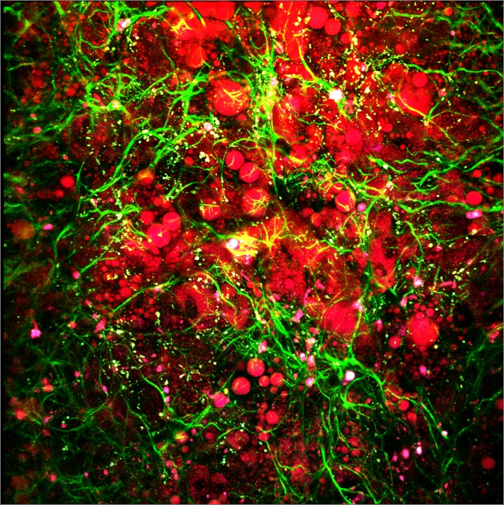
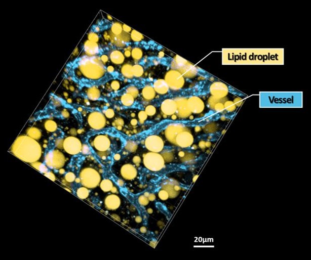
Lipid Droplet / Microvasculature visualized in vivo in 3D in the liver of Non-Alcoholic Fatty Liver Disease (NAFLD) mouse model
Liver Fibrosis visualzied in vivo by Second Harmonic Generation (SHG) Imaging in live nonalcholic fatty liver mouse model
Operation of custom-design, video-rate, intravital confocal microscopy system; starred by K. Choe
We are focusing on the development of the novel in vivo visualization techniques for the live animal in a single cell to single molecule resolution. In a recent decade, rapid advances of in vivo micro-visualization technologies have allowed us to catch a glimpse of numerous exciting processes such as gene expression, regulation, protein activity, drug delivery, cell trafficking, cell-cell interaction, physiological response to external stimuli in the natural microenvironment in vivo. We're now on the verge of a new era of micro-nano-scale in vivo visualization technologies those can be utilized as a novel versatile tool for basic and translational biomedical research as well as a valuable clinical tool for cellular-level diagnosis and monitoring.
Our ultimate goal is to open up a new avenue to answer questions in biomedical science those are important but difficult to investigate by pioneering innovative visualization technology. We are looking for a talented student and researcher in multiple disciplines including engineering, physics, chemistry, biology and medicine as an active interdisciplinary team-work is the cornerstone to accomplish our goal.
Positions open for graduate student (MS/PhD), Post-doctoral researcher [email]

[Award] Jieun Choi, awarded Best Poster Award, Advanced Biophotonics Conference (ABC), Jeju,
Nov. Korea, 2023
[Award] Lucia Stephani Edwina, awarded Best Poster Award, Korean Society for Vascular Biology
and Medicine (KVBM) Annual Meeting, Busan, Korea, Nov. 2022
[Award] Jieun Choi, awarded Best Poster Award, Advanced Biophotonics Conference (ABC), Pohang,
Nov. Korea, 2022
[Award] Lucia Stephani Edwina, awarded Best Paper Award, Optical Society of Korea (OSK),
Feb. Korea, 2022
Nature Methods, 20 (10), 1581-1592 (2023)
Chemical Communications, 59 (67), 10109-10112 (2023)
Detection of Retinal and Choroidal Neovascularization, Cells, 12 (14), 1902 (2023)
support associative social memory in male mice, Nature Communications, 14 (1), 2597 (2023)
as a Wound Healing Dressing, ACS Applied Materials & Interfaces, 15 (15), 18653-18662 (2023)
[Paper] In vivo longitudinal 920 nm two-photon intravital kidney imaging of a dynamic 2,8-DHA crystal
Biomedical Optics Express, 14 (4), 1647-1658 (2023)
the glioblastoma invasion zone, Experimental & Molecular Medicine, 55 (2), 470-484 (2023)
nitroaromatic-sensitive fluorescence, Polymer, 265, 125577 (2023)
J. Clinical investigation, 32 (24), e159672 (2022)
[Paper] Hematopoietic stem and progenitor cells integrate microbial signals to promote post-inflammation
gut tissue repair, The EMBO Journal, 41 (22), e110712 (2022)
[Paper] Two distinct receptor-binding domains of human glycyl-tRNA synthetase 1 displayed on extracellular
vesicles activate M1 polarization and phagocytic bridging of macrophages to cancer cells, Cancer Letters, 539, 215698 (2022)
Glia, 70 (5), 975-988 (2022)
Biomedical Optics Express, 13 (8), 4160-4174 (2022)

Welcome to In Vivo Micro-Visualization Lab.
What's New
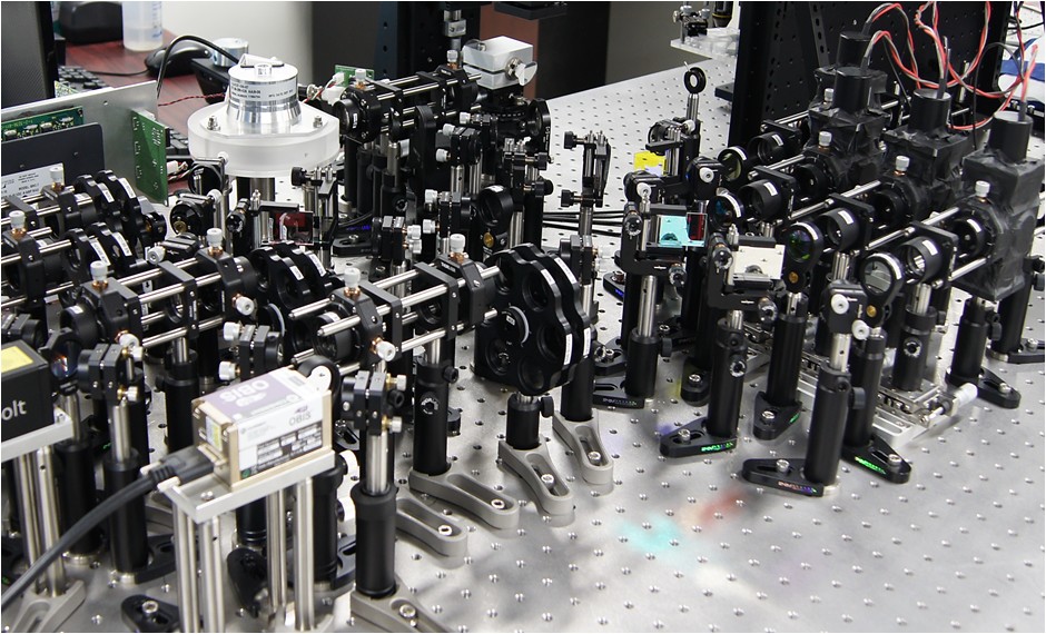
Custom-built Intravital
Confocal Microscopy
In Vivo Micro-Visualization Laboratory
Graduate School of Medical Science and Engineering (GSMSE)
Korea Advanced Institute of Science and Technology (KAIST)

In Vivo Micro-Visualization Laboratory
Graduate School of Medical Science and Engineering, Korea Advanced Institute of Science and Technology (KAIST)
Rm.219, Basic Research Building (E6-6), 291 Daehak-ro, Yuseong, Daejeon, 34141, Republic of Korea

Last update. 2024. 04. 08.